Everyone in my class just recently had the good fortune to be able to do a dissection, an incredibly useful and interesting opportunity. My group got the opportunity to dissect a cow's knee, which was absolutely fascinating. Getting a chance, as a
design student to tear apart an animal joint to look at the way it works and use this knowledge for your designs is an opportunity I highly doubt I'll ever get to have again.
Warning, all of these photos following show the cow's knee being dissected. If you are a little squeamish, I recommend skipping on to the next blog post.
The parts that I found the most fascinating were the way the tendons bound the whole knee together, and the way they weren't attach, but were
fused to the bone. It's impossible to distinguish the two of them. It's (coming from a design background, so do
not take my words as the reality; these are merely my limited understanding) as if there is no boundary between the tendon and the bone. They meld in the way that few things do.
The cow knee was given to us sawn off above and below the knee, and at first it was not immediately clear which side of the joint was the front of the knee. Bending the knee was impossible, so that also didn't give any hints. We had to delve deeper into the structure before any understanding was possible.
Some of the most fascinating stuff was the smoothness of the bone, and the way the two bones fit so perfectly together. The knee cap, seen top right, fits and slides beautifully (yes,
beautifully) along the upper knee bone and had extensive tendons and sinews connecting it to the surrounding tissue and the other bones.
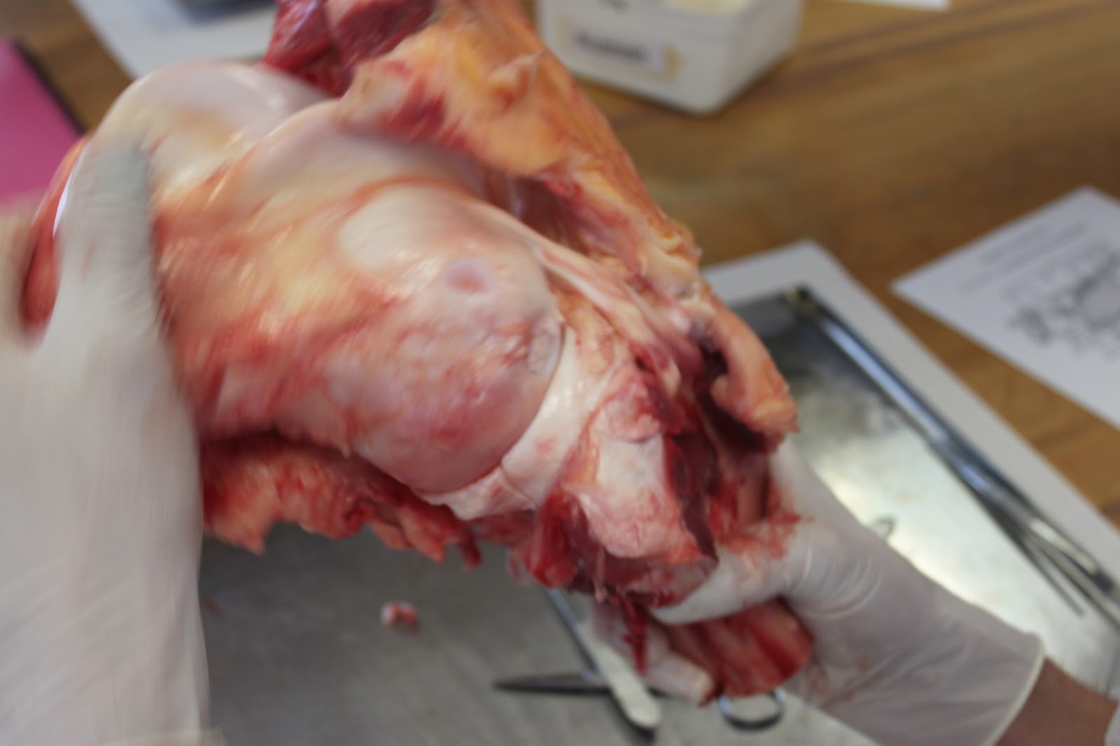
Once I had severed some of the constraining tissue, I was able to manipulate the joint as shown above. What we see is the tissue containing the knee cap and fat sliding across the upper knee bone, the
femur (assuming the bone is called the same thing for cows). It's not immediately clear what the knee cap is there for, however it looked like it was re-directing some of the force and impact of the weight around the joint, as well as protecting it.
Another excellent example of fantastically different tissue was the cartilage that sits in between the femur and tibia, which formed a disc that essentially provided the lubricant and the shock-absorber for the joint. Another thing we learnt was that the blood diffuses from the surrounding tissue into the cartilage, because the cartilage doesn't have any blood vessels of its own.
So, overall, a very interesting dissection, providing very valuable insights for the second project. Updates on that coming soon!


No comments:
Post a Comment